| Purpura_fulminans = الفرفرية الصاعقة |
|
| atlas of dermatology - P | |||
| Friday, 22 October 2010 18:33 | |||
|
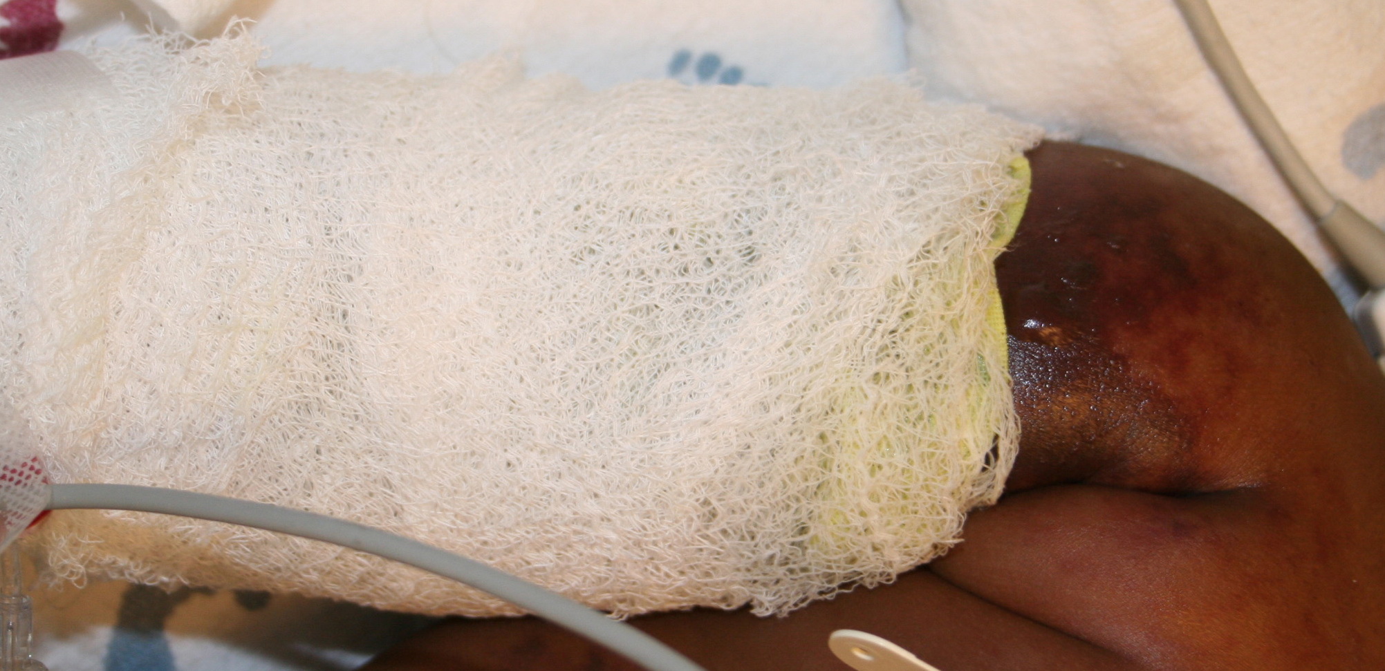
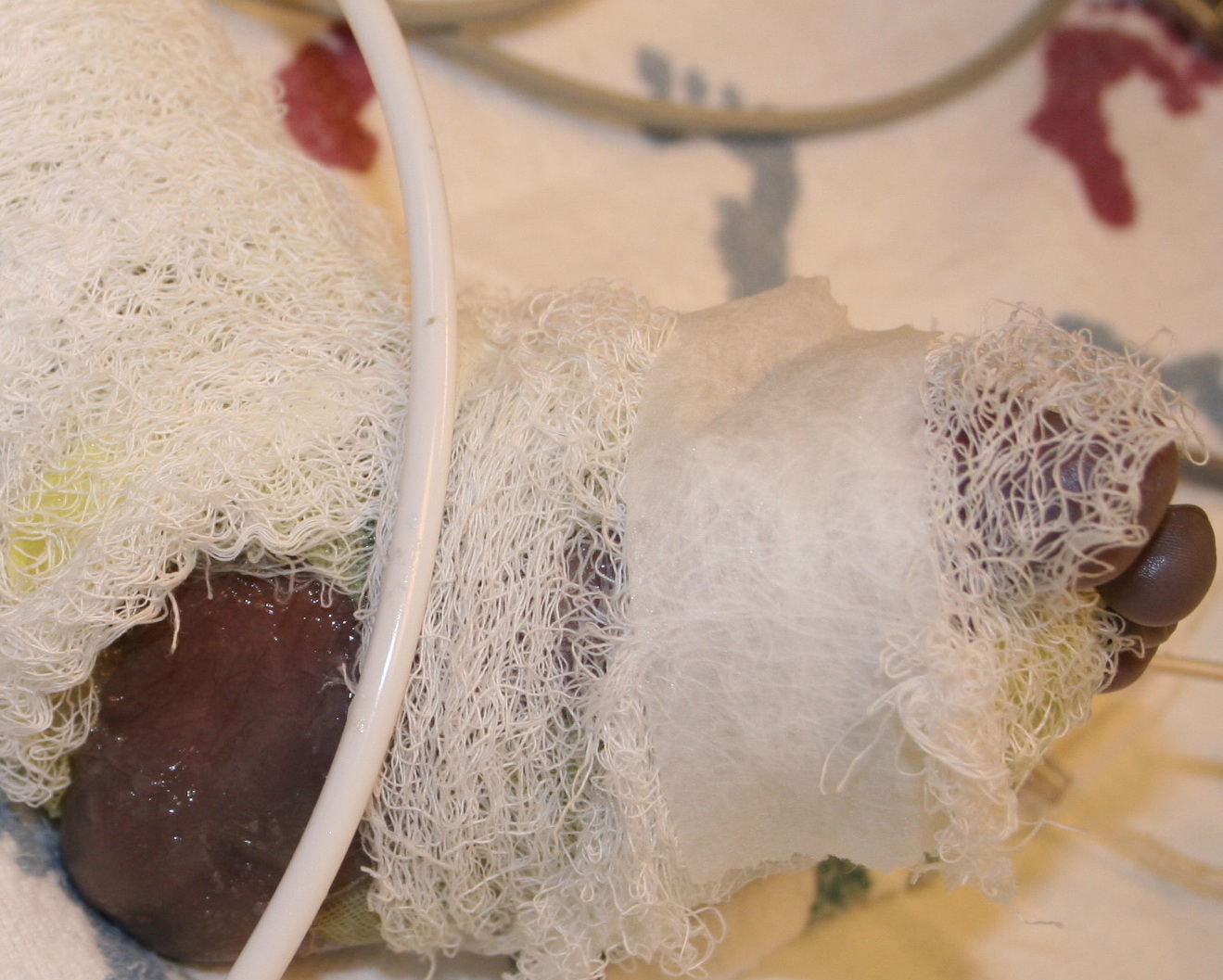
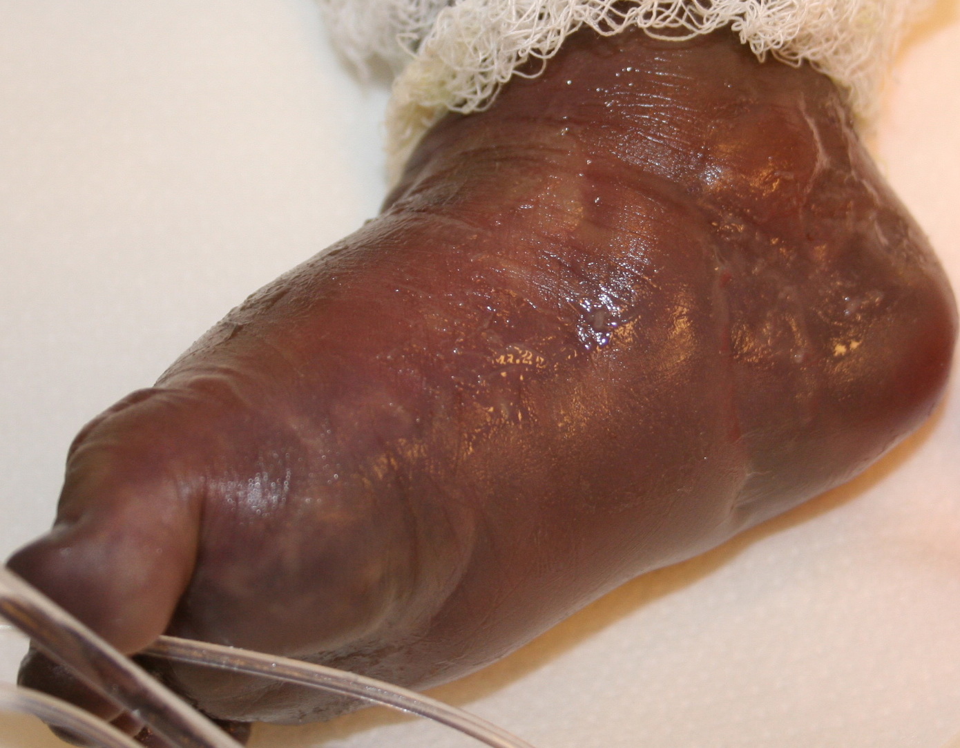
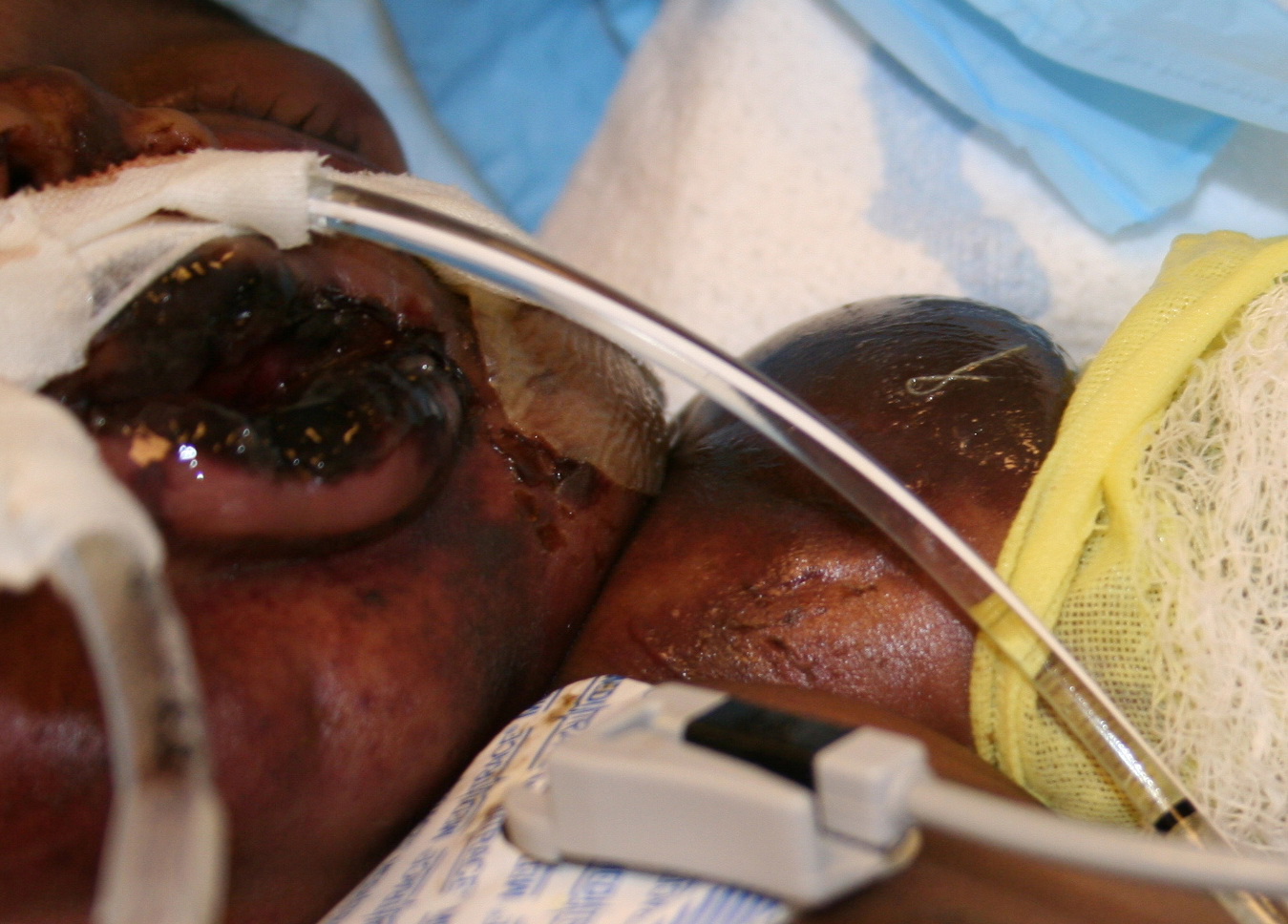
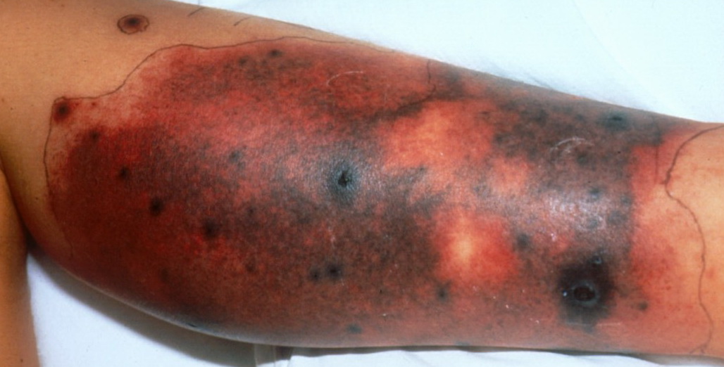
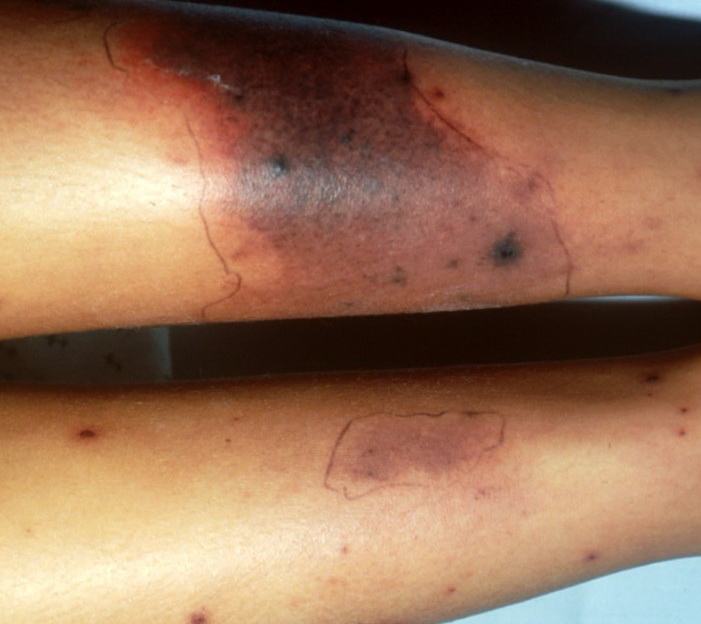
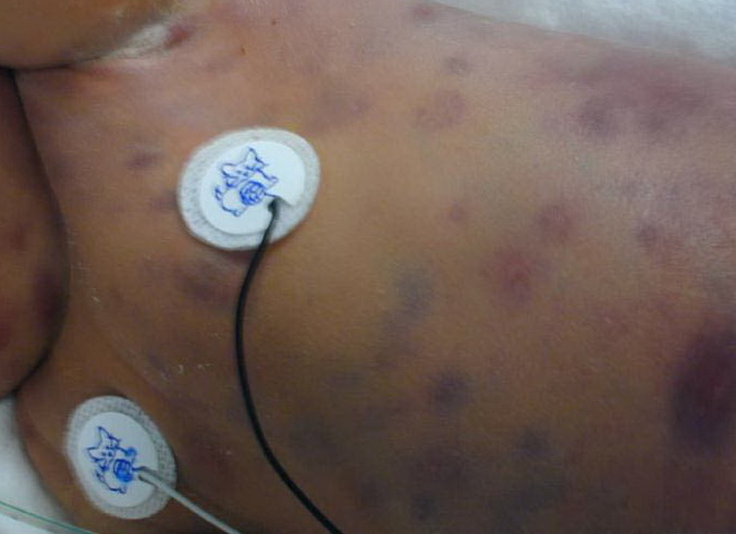
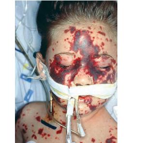
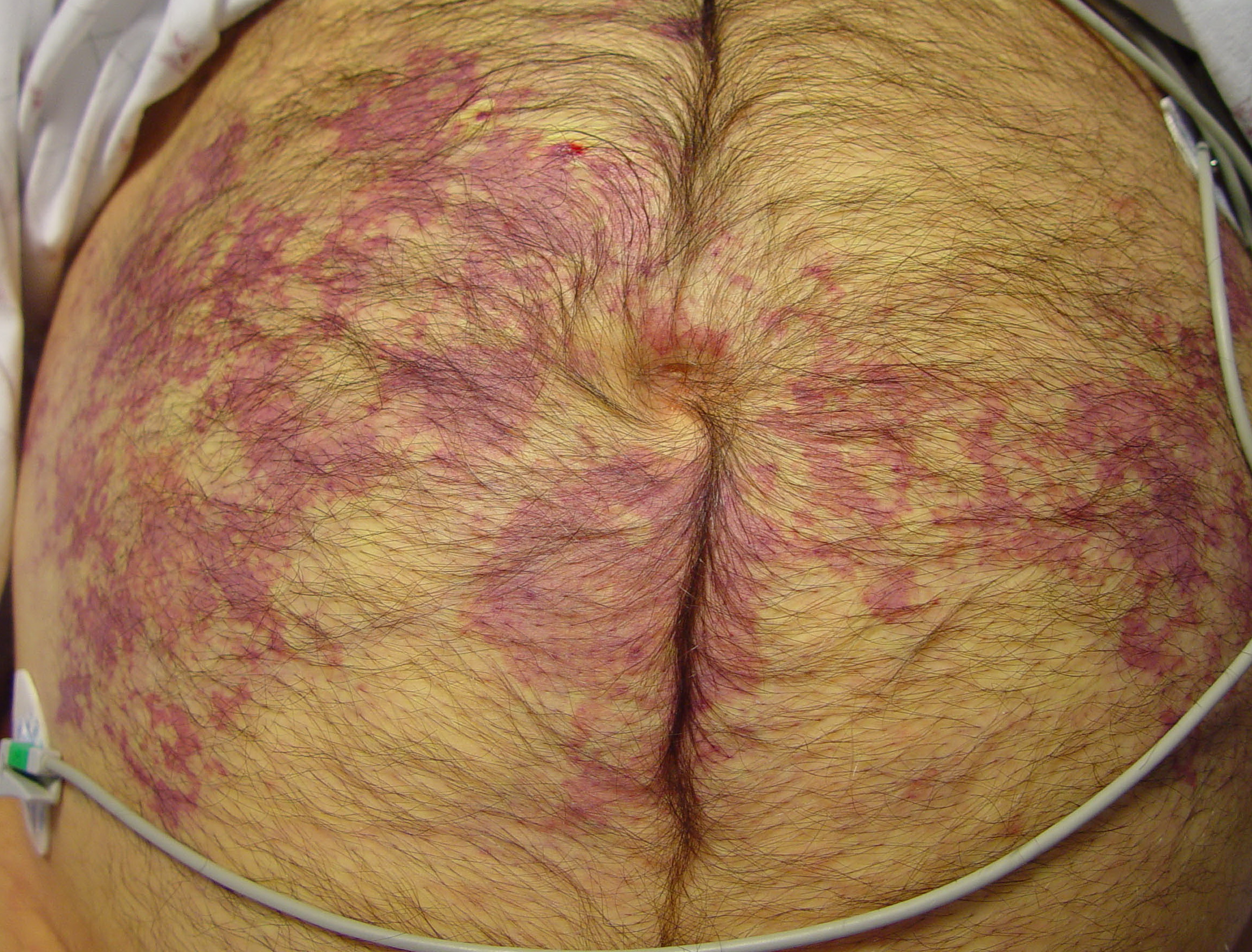
PURPURA FULMINANS
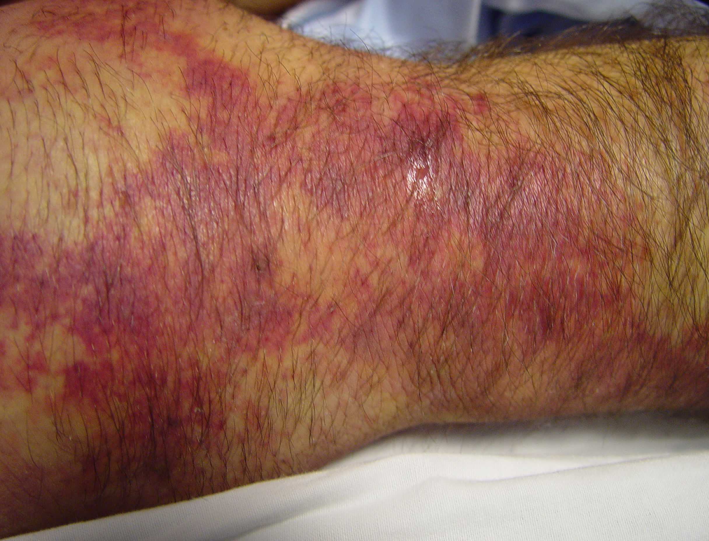
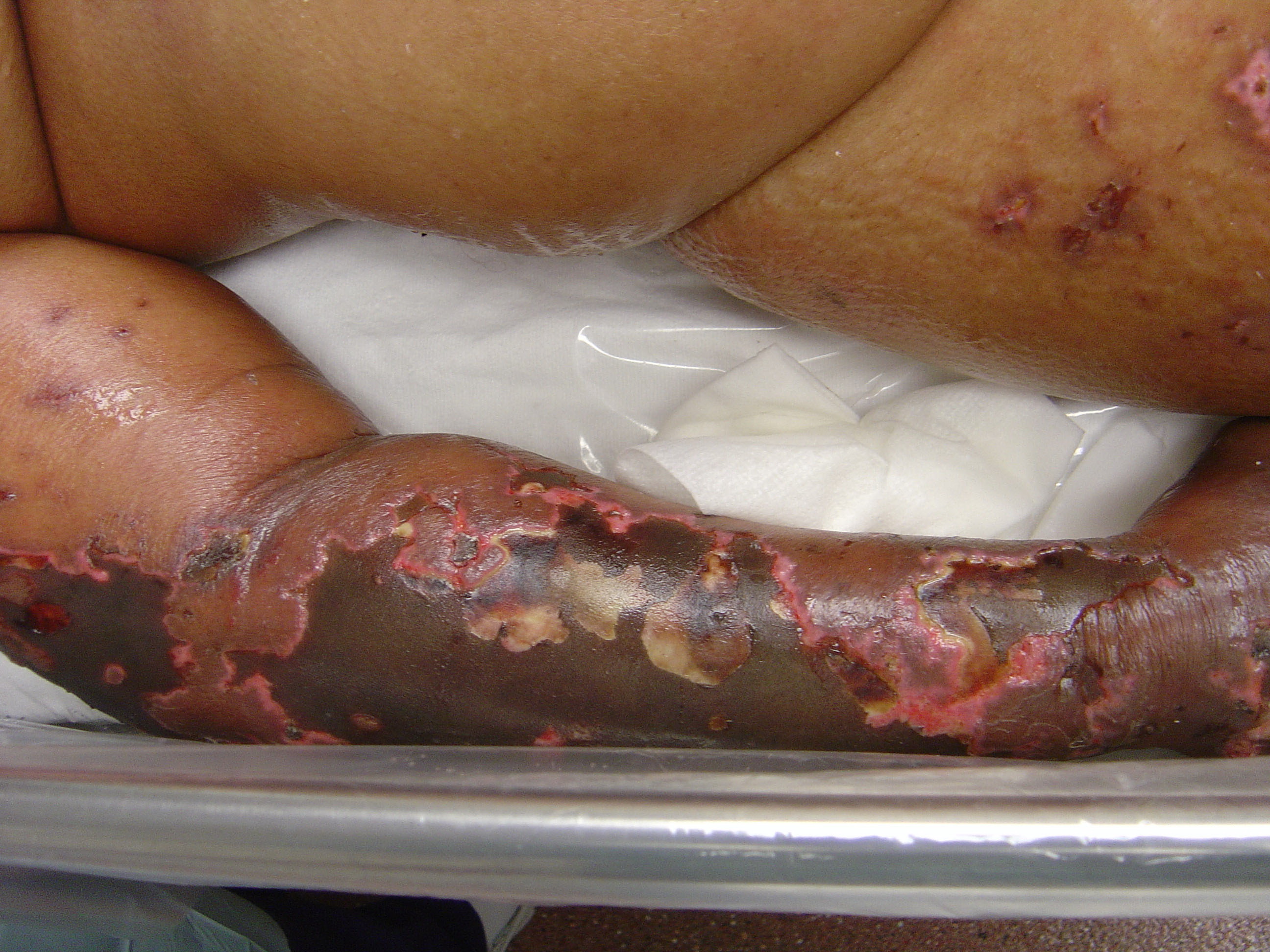
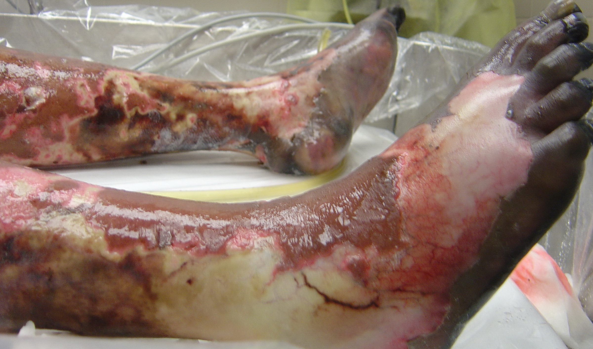
Purpura fulminans is a severe skin disorder associated with DIC that mainly affects children and infants. Extensive areas of skin develop blue-black hemorrhagic necrosis, and biopsy typically reveals small-vessel microthrombi and vasculitis. Distribution of necrotic areas typically depends on the clinical setting. Patients are acutely ill with fever and hemorrhage from multiple sites, are hypotensive, and manifest typical laboratory signs of DIC. The pathogenesis of purpura fulminans is unknown, but histologic findings have been likened to the animal model of consumptive coagulopathy, the local Shwartzman reaction. It has also been suggested that the development of purpura fulminans in meningococcemia may result from acquired defects in the protein C pathway because similar lesions are seen in two other protein C deficiency states, namely, neonatal purpura fulminans and warfarin-induced skin necrosis.
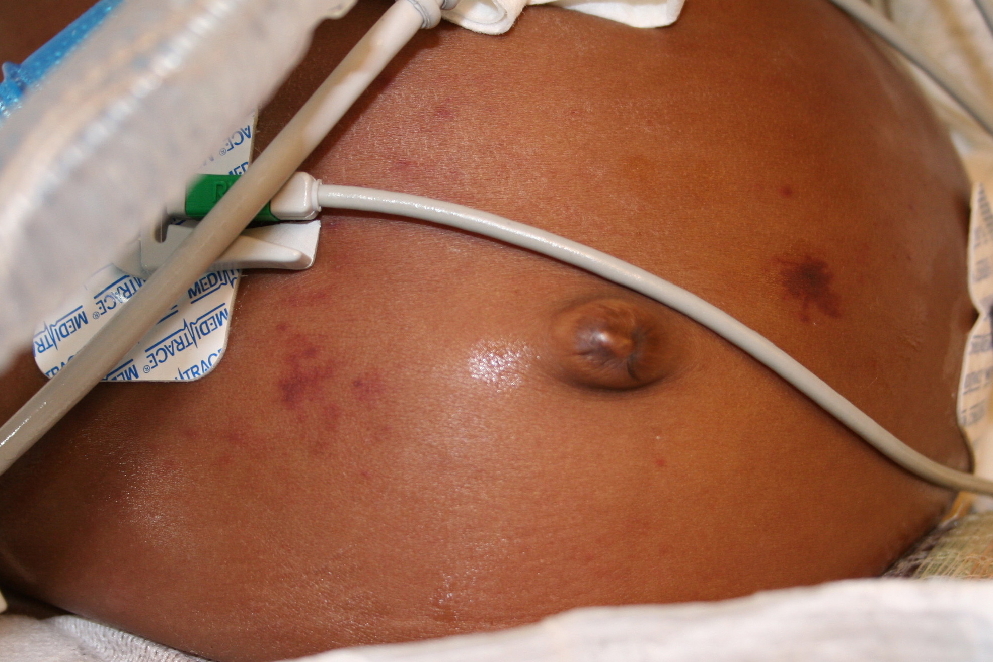
Purpura fulminans occurs in three clinical settings. In acute sepsis-associated or secondary purpura fulminans, the initial presentation is one of overwhelming infection that is most commonly meningococcemia but has been reported with many other Gram-positive and Gram-negative organisms. Patients are hypotensive with reduced peripheral perfusion contributing to skin necrosis on distal extremities and elsewhere ; alternatively, skin necrosis can occur in a patchy distribution. Idiopathic purpura fulminans can manifest within 10 days of an antecedent illness, most commonly scarlet fever and varicella in children. Most skin lesions are localized to the breasts or lower half of the body. Third, purpura fulminans can be seen in association with homozygous protein C deficiency in affected neonates, in whom massive intravascular thrombosis and abdominal wall gangrene can also develop. Acquired inhibitors of the protein C pathway can also be associated with this presentation. Patients with warfarin-induced skin necrosis may present with a similar but usually less severe form of purpura fulminans and warrant consideration of protein C or protein S deficiency. The mortality rate has recently been significantly reduced in purpura fulminans, largely because of more widespread use of therapeutic heparinization in these patients and aggressive transfusion support factor replacement.
DISSEMINATED INTRAVASCULAR COAGULATION DIC is a syndrome characterized by a systemic activation of coagulation leading to the intravascular deposition of fibrin in the (micro) vasculature and the simultaneous consumption of coagulation factors and platelets.The occurrence of microvascular thrombosis as a consequence of DIC is underscored by pathologic and clinical findings, demonstrating a link between DIC and organ dysfunction. Pathogenetic pathways that play a role in the development of DIC include tissue factor-dependent activation of coagulation, defective physiologic anticoagulant pathways (such as the antithrombin system and the protein C system), and impaired fibrinolysis, caused by elevated levels of plasminogen activator inhibitor type 1. New therapeutic strategies are based on current insights into the pathogenesis of DIC, and include anticoagulant strategies (e.g., directed at tissue factor) and strategies to restore physiologic anticoagulant pathways (such as activated protein C concentrate). DIC is not a disease in itself but is always secondary to an underlying disorder. Hyperglobulinemic purpura of Waldenström is characterized by recurring nonthrombocytopenic purpura, hypergammaglobulinemia, and an increased sedimentation rate. Hyperglobulinemic purpura of Waldenström may present alone as a primary entity or as a secondary form associated with diseases, such as Sjögren syndrome or systemic lupus erythematosus. Multiple therapies including corticosteroids, antimalarials, plasmapheresis, and other anti-inflammatory and/or immunosuppressive agents have led to variable results. The cutaneous lesions associated with scurvy are characterized by purpura that surrounds corkscrew hairs on the lower extremities . Senile purpura results from the loss of elasticity of the skin and weakened capillary walls. RBC extravasation occurs with minor trauma to the hands and other sites Solar purpura usually occurs on the extensor surfaces of the forearm. The pigmented purpuric dermatoses are discussed in detail in Chapter 169.
|
|||
| Last Updated on Friday, 12 November 2010 08:47 |